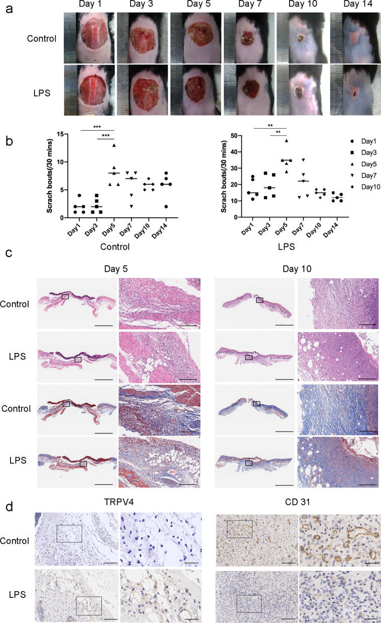
Fig. 1.
Application of LPS-induced delayed wound healing with itching. a Representative photographs showing macroscopic excisional wound closure of C57BL6 mice subcutaneously injected with PBS (Control), or 0.5 mg/kg LPS (LPS). b Itching behaviors of mice with cutaneous wounds during wound healing. n = 5. *P < 0.05; **P < 0.01; ***P < 0.001. c Representative H&E and Masson-stained sections of PBS- or LPS-injected incisional wounds. Scale bars (low power field), 3 mm. Scale bars (high power field), 200 µm. d Representative immunohistochemistry of TRPV4 on day 5 and CD31 on day 10 within PBS- or LPS-injected incisional wounds. Scale bars (low power field), 100 µm. Scale bars (high power field), 30 µm

