Fig. 1.
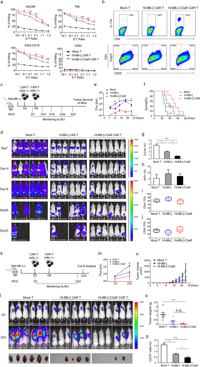
19-BB-ζ-C3aR CAR-T cells displayed potent anti-leukemia activity in vitro and in vivo, particularly in the xenografts extramedullary leukemia model. a The 19-BB-ζ-C3aR CAR-T cells showed significantly increased ability to lyse CD19-expressing tumor cells compared to 19-BB-ζ CAR-T cells. The cytotoxicity assay was performed at least three independent experiments. b Flow-cytometry results revealed enhanced expansion of Th17 cells and reduced Tregs in the 19-BB-ζ-C3aR CAR-T cells compared to 19-BB-ζ and mock-transduced T cells. c To establish the ALL model, 5 × 10⁵ NAML6-Luc cells were administered intravenously into NCG mice, which were randomized to the treatment with 2 × 10⁶ indicated T cell on Day 2 and Day 8. NAML6 tumor growth was then monitored by Xenogen imaging. d Bioluminescence images of NCG mice at Days 7, 14, 19, 26, and 33 are depicted for each group. e The curve of flux on indicated time points. f Kaplan–Meier survival analysis for ALL mice. Log-rank tests were used to perform statistical analyses of survival between groups. g The 19-BB-ζ-C3aR CAR-T group showed significantly fewer blast counts than Mock and 19-BB-ζ CAR-T groups. h The detectable GFP-positive T cells were similar in three groups. i, j There were no differences in CD4+ and CD8+ T cells between the indicated T cell populations. k Xenograft extramedullary leukemic model was established by subcutaneous injection of 5 × 10⁵ NALM-6 cells. The indicated CAR-T with 2 × 10⁶ dose were intravenously injected on Day 3 and Day 8, respectively. l NAML6 subcutaneous tumor growth was monitored by Xenogen imaging. m The curve of flux on indicated time points. n, o The tumor mass and weight were measured and recorded. p The 19-BB-ζ-C3aR CAR-T group showed the lowest CD19+ ALL blast counts on Day 22. ***p ≤ 0.001; **p ≤ 0.01; *p ≤ 0.05, n.s. no significant

