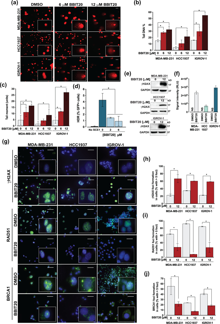
FIGURE 5.
BBIT20 enhances DNA damage by inhibiting homologous DNA repair, in triple-negative breast and ovarian cancer cells. (a–c) Measurement of DNA damage in cancer cells, after 48 h of treatment with 6 and 12 μM BBIT20, was measured by comet assay. In (a), representative images are shown (scale bar = 20 μm; 200X magnification). In (b), quantification of tail DNA percentage; in (c), quantification of tail moment; data are mean ± SD, n = 5 independent experiments (200 cells per sample); *P <0.05 significantly different from DMSO (two-way ANOVA followed by Dunnett’s test). (d) Homologous DNA repair activity was determined in MCF7 DR-GFP cells treated with 6 and 12 μM BBIT20, using the chromosomal DR-GFP assay; data are mean ± SD, n = 5 independent experiments; *P <0.05 significantly different from DMSO (one-way ANOVA followed by Dunnett’s test). (e, f) Analysis of γH2AX expression levels in cancer cells, after 48 h of treatment with 12 μM BBIT20. In (e), representative immunoblots are shown; GAPDH was used as loading control. In (f), quantification of protein expression levels relative to DMSO; data are mean ± SD, n = 5 independent experiments; RLU (relative light unit). (g) γH2AX, RAD51 and BRCA1 foci formation, and BRCA1 cellular localization after 48 h of treatment with 12 μM BBIT20; representative images (scale bar = 100 μm; 400X magnification) are shown. Quantification of γH2AX (h), RAD51 (i) and BRCA1 (j) foci formation; data are mean ± SD, n = 5 independent experiments (100 cells per sample); *P <0.05 significantly different from DMSO (unpaired Student’s t-test)

