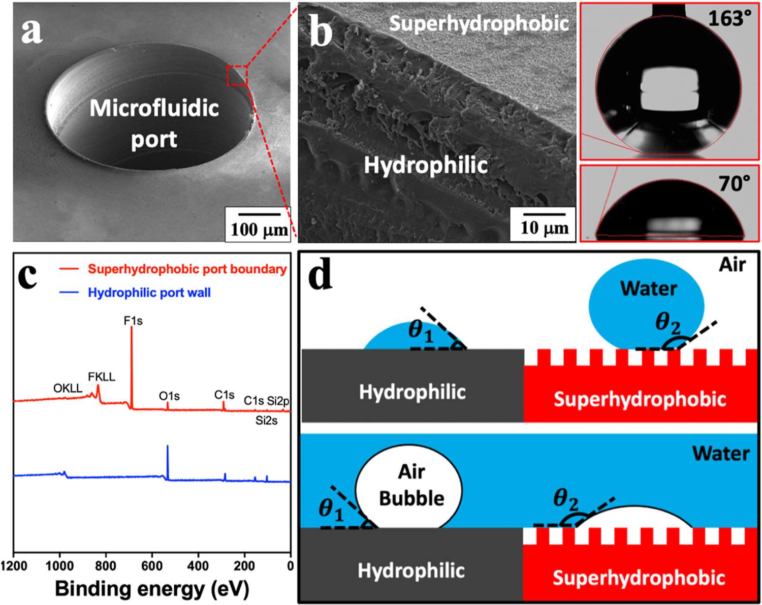
Fig. 2.

Sharp wettability contrast at the microfluidic port edge. (a) SEM image of a microfluidic port. (b) Close-up image shows the edge of the microfluidic port and the static water CAs before/after superhydrophobic silica coating. (c) XPS analysis on superhydrophobic port peripheral and hydrophilic inner port wall. (d) Schematics showing the interaction of in-air water droplets and underwater air bubbles with hydrophilic and superhydrophobic surfaces, respectively.
