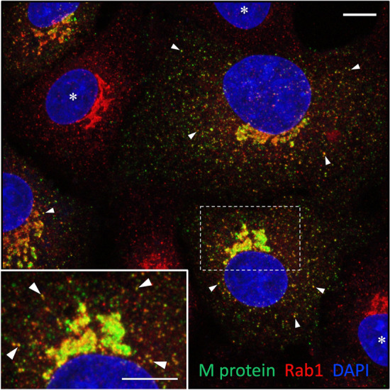
Fig. 2.

The IBV M-protein colocalizes extensively with IC marker Rab1. The IBV-infected cells were fixed at 8 hpi and double-stained for CM using antibodies against Rab1 and the M protein, which predominantly associates with intralumenal virus particles. Note the variable expression of the M protein in different virus-infected cells. The inset highlights the Golgi region of a cell with strong M expression, leading to the development of membrane domains that evidently contain progeny virus, but are devoid of Rab1. Peripheral IC elements positive for both M and Rab1 are indicated by arrowheads, while uninfected cells are denoted by asterisks. The nuclei were visualized by DAPI staining. Bars, 5 μm
