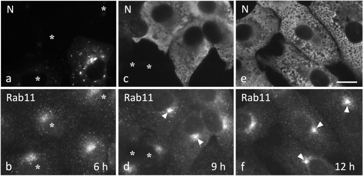
Fig. 4.
IBV infection of Vero cells results in ERC compaction. The cells fixed at 6, 9, or 12 hpi were double-stained for immunofluorescence microscopy with antibodies against Rab11 and the viral N protein—to localize REs and identify infected and uninfected (asterisks) cells, respectively. Note the gradual compaction of the Rab11-positive pericentrosomal ERC (arrowheads) in the course of the infection and simultaneous reduction of the peripheral Rab11 signal. Bar, 10 μm

