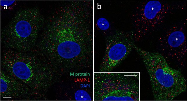
Fig. 8.

Non-overlapping localizations of the M protein and LAMP-1 in IBV-infected Vero cells. The cells fixed at 8 (a) or 12 hpi (b) were double-stained for the viral M protein and LAMP-1, and subjected to imaging by CM. Despite their strong expression and joint accumulation in the perinuclear Golgi region in part of the cells (see b, inset), the proteins display negligible colocalization. Also note the similar LAMP-1 staining patterns of the infected (M-positive) and uninfected (M-negative; b, asterisks) cells. Bars, 5 μm
