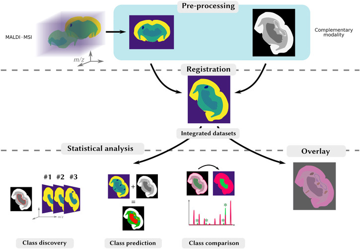
FIGURE 1.
Typical processing and analysis workflow for multimodal MALDI-MSI. Top-row: the MALDI image (on the left), and the complementary images (on the right) are processed so as to have comparable shapes, through various specific processing steps, including segmentation. Middle-row: the images are spatially aligned through registration. Bottom-row: once the images are registered, they can be visualized on-top of each other to reveal similar spatial distributions (on the right). Patterns can be objectified by a joint statistical analysis to, e.g., find spatial correlations, by investigating spatial clusters (class discovery), produce an enriched dataset from both modalities (class prediction) or find region-specific ions in two different ROIs (class comparison).

