
Fig. 4.
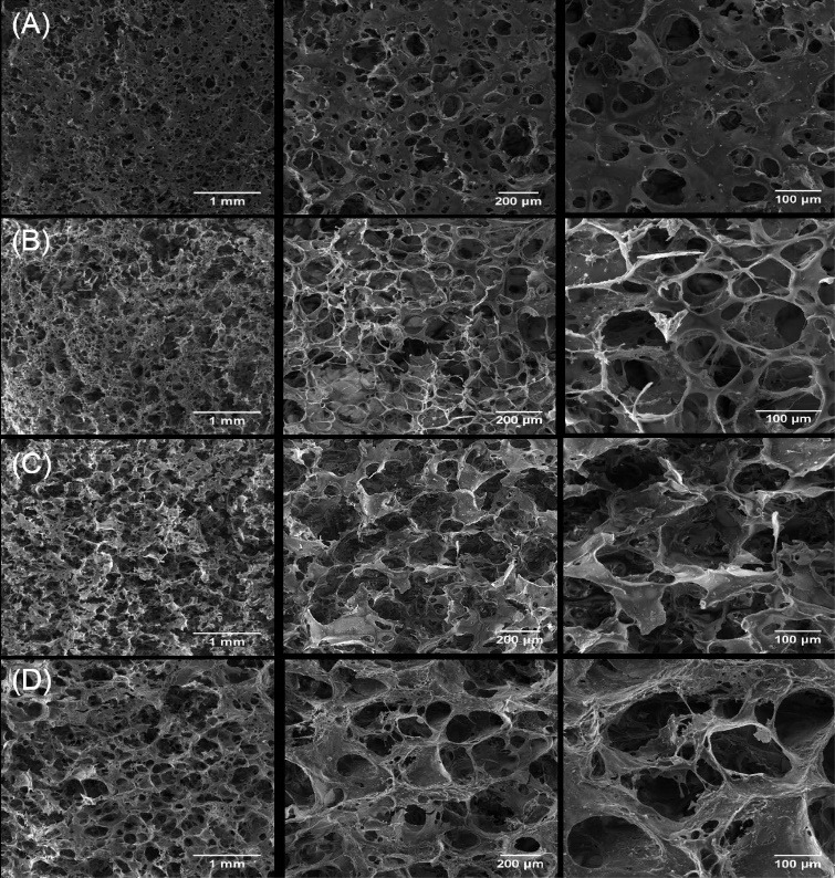
FE-SEM micrographs of pure and Cu-substituted nHA/Cs/Gel scaffolds Monitoring the scaffolds by FE-SEM verified the microscopic spongy and highly porous architecture in which pores were connected to make an interconnected 3-dimensional network. The micropores were found in a variety of sizes mainly < 100 μm for nHA/Cs/Gel scaffolds and > 100 μm for Cu incorporated scaffolds.