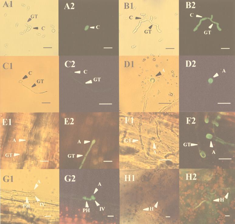
FIG. 1.
Developmental expression of gfp under the control of the clpg2 promoter. Conidia of C. lindemuthianum H2 were allowed to germinate in vitro either on pectin medium cleared by filtration (A and B) or on a glass slide in water (C and D). They were also used to inoculate bean hypocotyls (E through H). Samples shown in panels A through D were assayed for green fluorescence after 12 h (panels A and C) and 24 h (panels B and D). Infected bean hypocotyls were examined 24 h (panels E and F), 48 h (panel G), and 15 days (panel H) after inoculation. Samples were successively analyzed by light microscopy (subpanels 1) or fluorescent light (subpanels 2). A, appressorium; C, conidium; GT, germ tube; IV, infection vesicle; PH, primary hyphae. Bar = 20 μm.

