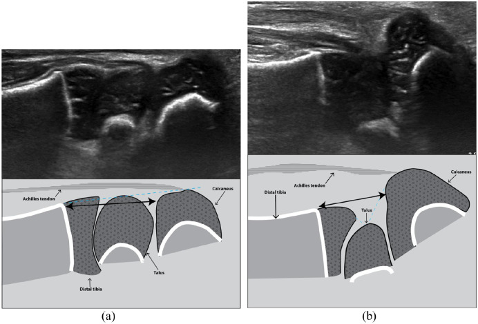
Figure 3.
(a) Posterior sagittal view in a normal foot. There is a normal alignment of the posterior aspect of the distal tibia, talus, and calcaneus (straight dotted blue line) and an appropriate distance between the distal tibial physis and the proximal calcaneal apophysis (Ti-C) (double black arrowhead). (b) Posterior sagittal view in a clubfoot. This view reflects the calcaneal position in the heel. The tibio-talo-calcaneal alignment is disrupted (broken dotted blue line) and the distance between the distal tibial physis and the proximal calcaneal apophysis (Ti-C) is decreased (double black arrowhead).

