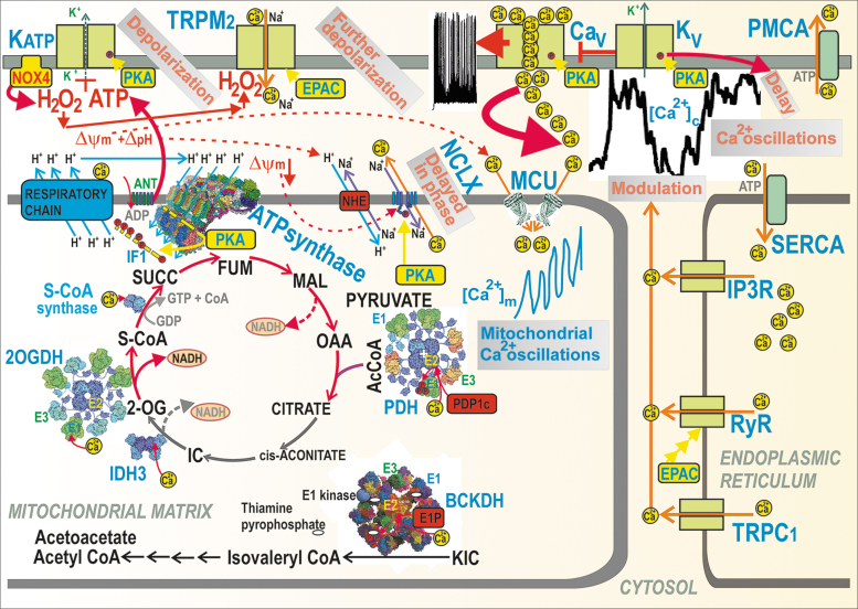
FIG. 4.
Mitochondrial versus cytosolic Ca2+ oscillations and their ability to activate matrix dehydrogenases. Action potential firing is reflected by cytosolic Ca2+ oscillations, which determine the steady-state increase in matrix [Ca2+]m with superimposed [Ca2+]m oscillations and concomitant activation of matrix dehydrogenases. Activation due to the PKA pathway is also indicated, demonstrating phosphorylation (red circles) of (i) IF1 hypothetically forming bridges (yellow) between the neighboring dimers of the ATP synthase within a row of dimers (four dimers are depicted with the indicated H+ backflow that leads to ATP synthesis); (ii) NCLX promoting activation via the ΔΨm decrease; (iii) KATP channels (setting their sensing of ATP to ∼1 mM [ATP]); (iv) CaV channels, thus activating them. Similarly, the EPAC2A pathway (“EPAC”) reportedly activates TRPM2 and RyR. The dashed arrows, pointing to NADH, illustrate the sites where NAD+ is made from NADH due to pyruvate redox shuttles (Fig. 5). CaV, voltage-dependent Ca2+ channels; EPAC, exchange proteins directly activated by cAMP; FUM, fumarate; IC, isocitrate; MAL, malate; NCLX, mitochondrial sodium calcium exchanger; RyR, ryanodine receptor; SUCC, succinate.

