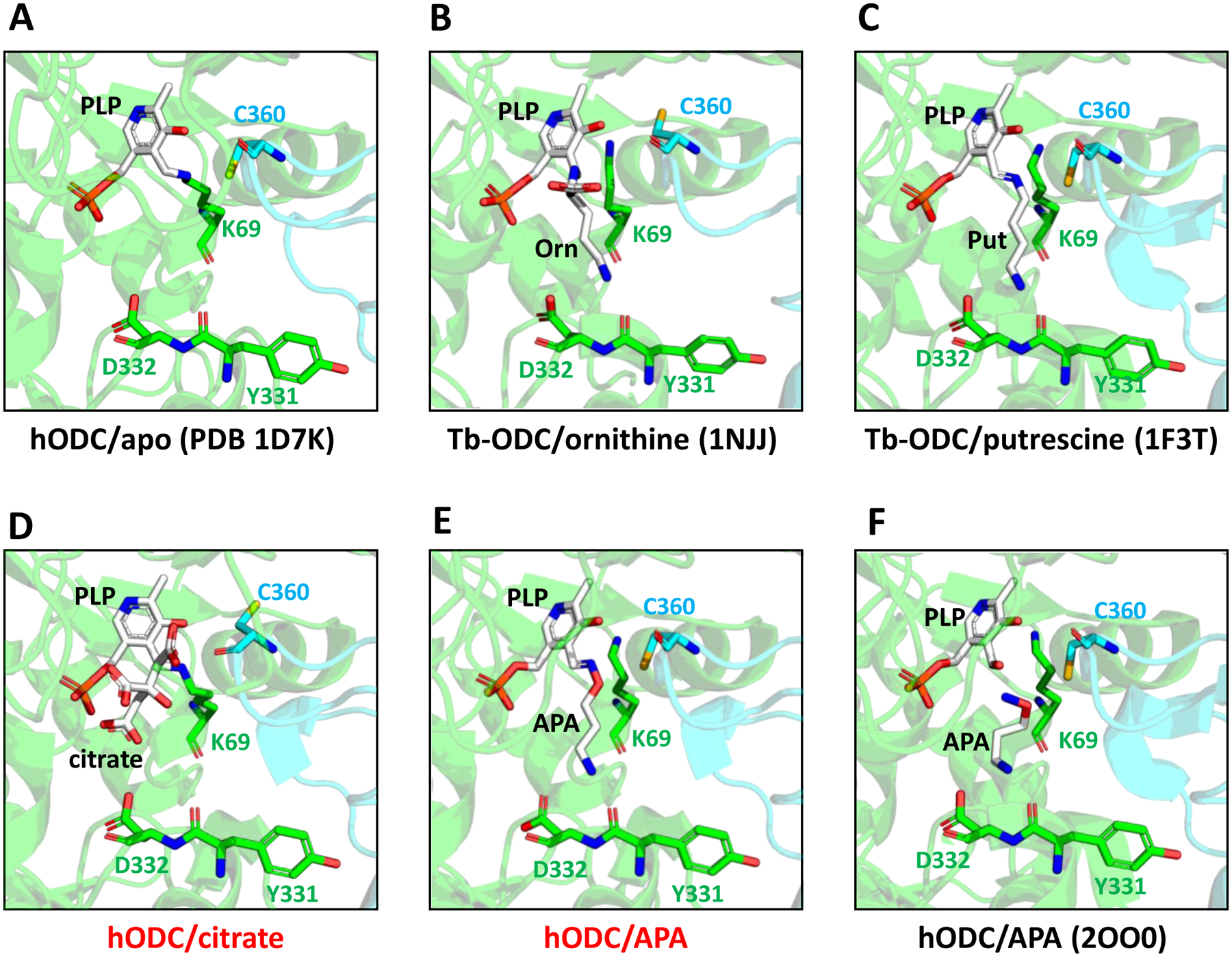
Figure 6: Structure close-ups of the catalytic center of different ODC complexes.

A. Human (h) apo-ODC. PLP (white) forms an internal aldimine with K69. The two monomers of the ODC homodimer are shown in green and cyan. B,C. Trypanosoma brucei (Tb) ODC bound to ornithine (B) and putrescine (C). D,E. hODC bound to citrate (D) and APA (E). F. Previously published structure (PDB 2OO0) of APA-bound hODC. PDB 2OO0 also contains cadaverine, but the position of cadaverine is outside of the binding pocket regions shown. Red letters: structures determined for this study; black letters: previously determined structures. Key residues are shown in stick representation.
