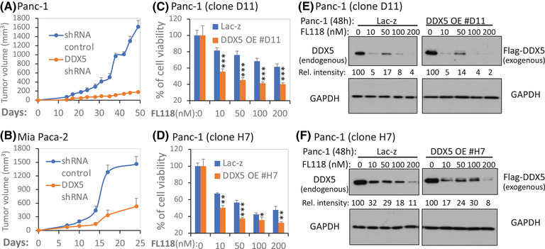
FIGURE 7.

Genetic modulation of DDX5 in PDAC cells affects tumour growth, cell viability and FL118 responsiveness. (A), (B) Silencing of DDX5 delays PDAC tumour growth: Control and DDX5‐specific shRNA lentiviral particle‐infected cells (2 × 10⁶) were subcutaneously injected into each site in the flank area of SCID mice. Tumour growth was monitored over time. The tumour growth curve from each time point is the mean ± SD from 5 tumours from five mice. (C), (D) Overexpression (OE) of DDX5 increases FL118 efficacy to inhibit PDAC cell growth/viability: Two DDX5 OE Panc‐1‐cell clones (C, clone #D11; D, clone #H7) in parallel with Lac‐z control Panc‐1‐cell clones were treated with and without FL118 as shown for 72 h. Cell viability was then determined using the MTT assay. The data are the mean ± SD derived from three tests. *p < .05; **p < .01; ***p < .001. (E), (F) FL118 could degrade both endogenous and exogenous DDX5. Panc‐1 D11 and H7 cloning cells that were forced to express Lac‐z (control) or Flag‐DDX5 were treated with and without FL118 treatment as shown for 48 h. Cells were then analysed using western blots with DDX5 antibodies (E, F, left panel) or with Flag antibodies (E, F, right panel). GAPDH was used as the internal control for total protein loading. The relative (Rel.) intensity of the western blot bands in each lane for DDX5 expression was provided by setting the band without FL118 treatment as 100 after being normalised to the GAPDH internal control
