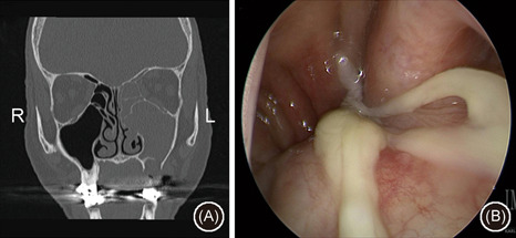
Figure 2.

Case example of utilization of maxillary antrostomy and anterior ethmoidectomy for a left‐sided odontogenic sinusitis; (A) coronal noncontrast CT scan demonstrating left‐sided maxillary and anterior ethmoid sinus opacification adjacent to a bony defect on the sinus floor with periapiacal abscess and (b) endoscopic exam demonstrating purulence emanating from the middle meatus and draining into the nasopharynx. CT, computed tomography
