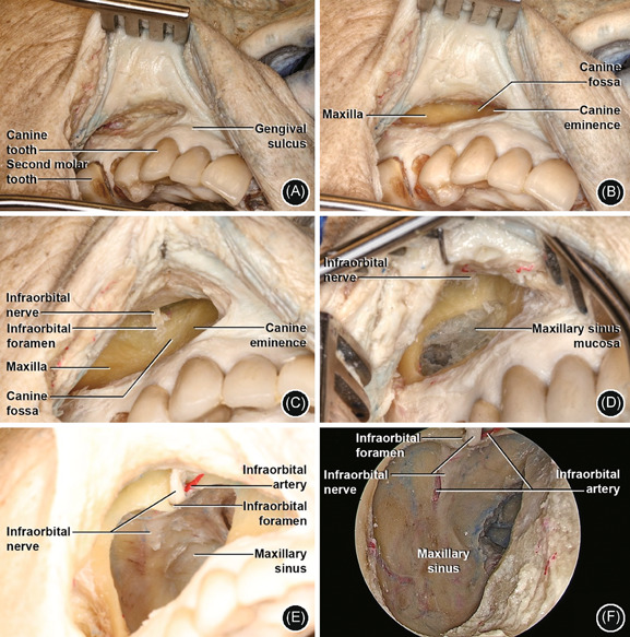
Figure 3.

Caldwell–Luc (anterior transmaxillary) approach. (A) Right side: incision between the canine tooth and second molar, about 5 mm above gingival sulcus; (B) detachment of periosteum of maxilla and identification of canine fossa and eminence inferior and medially; (C) identification of IOF and ION in the central‐superior area; (D) window in anterior wall (3 cm diameter) with visualization of maxillary sinus mucosa. ION in central‐superior area; (E) removal of sinus mucosa and opening into the MS cavity. Visualization of ION and IOA; (F) 4 mm 0‐degree endoscope MS anterior view with ION and IOA in the posterior wall. IOA, infraorbital artery; IOF, infraorbital foramen; ION, infraorbital nerve; MS, maxillary sinus
