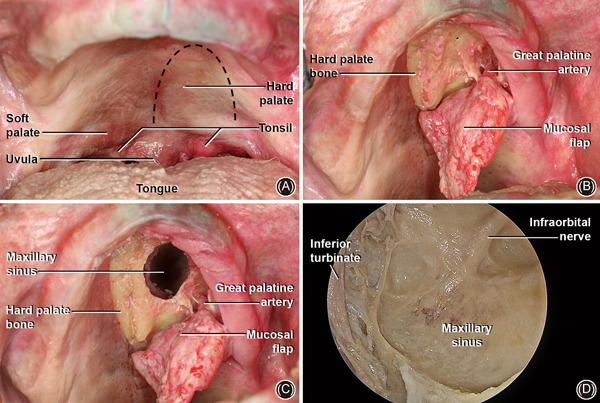
Figure 7.

Transpalatal approach. (A) Overview of the oral cavity with hard palate visualization and inverted “U” incision in left hemipalatal region. (B) Mucoperiosteal flap with hard palate bone exposure. Greater palatine artery in inferior/lateral portion of hard palate; (C) a burr hole has been performed in the hard palate with an inferior view of the MS, mucosal flap, and GPA; (D) 4 mm 0‐degree endoscopic inferior overview of MS with ION in the posterior wall. GPA, great palatine artery; ION, infraorbital nerve; MS, maxillary sinus
