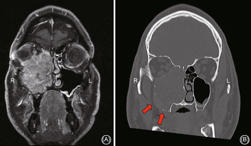
Figure 8.

Case example of a right‐sided maxillary sinus mucosal melanoma requiring a combined transpalatal and transfacial approach with orbital exenteration; (A) coronal T1 postgadolinium MRI scan showing intraorbital extension and (B) coronal noncontrast CT scan demonstrating bony erosion and tumor involvement along the floor of sinus necessitating transpalatal approach. CT, computed tomography; MRI, magnetic resonance imaging
