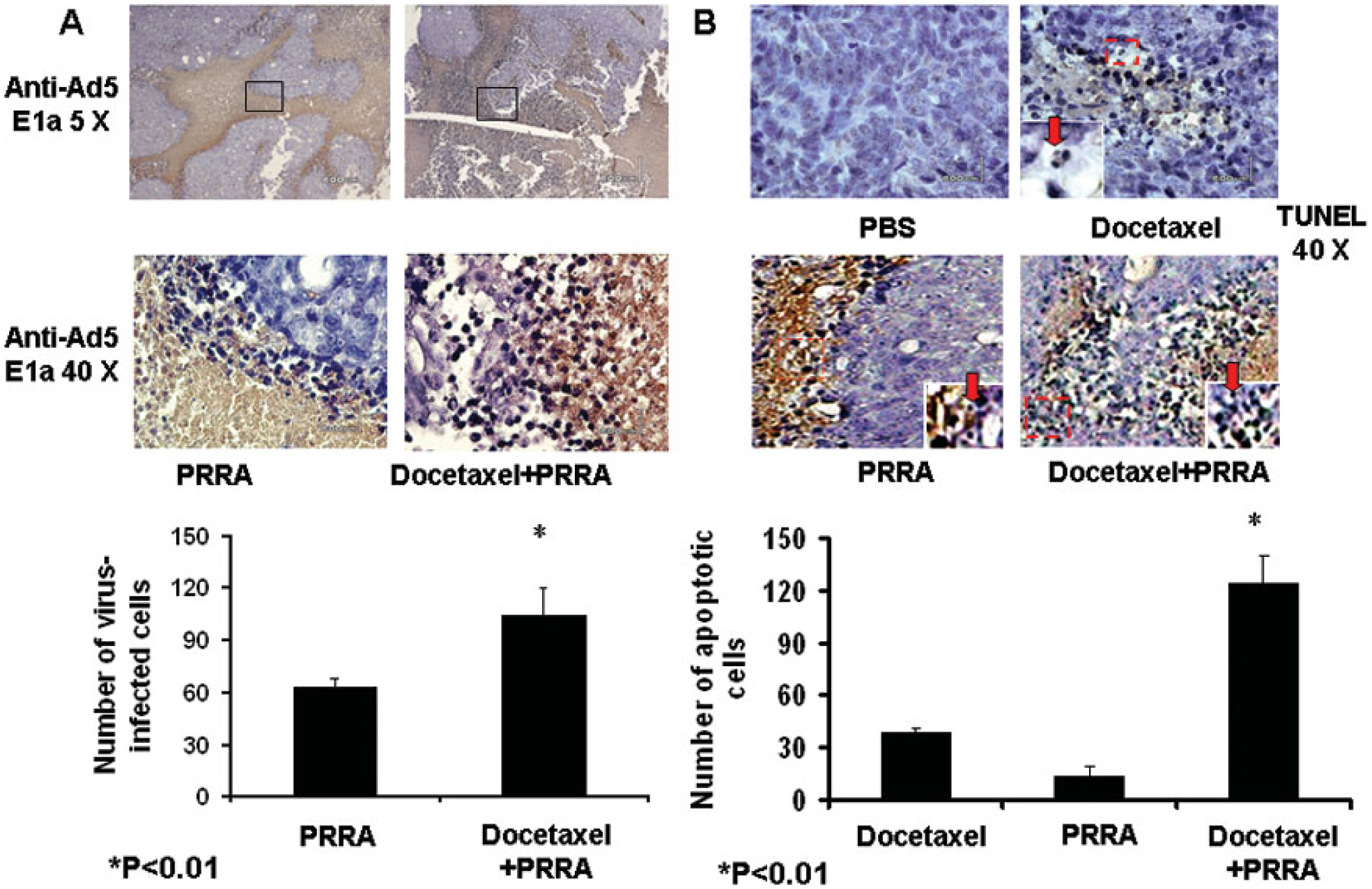
Fig.3.

Docetaxel significantly enhanced virus distribution and cell apoptosis. Tumors in PRRA-treated or docetaxel plus PRRA-treated mice were harvested after dual-photon imaging. A, Adenovirus distribution was evaluated by immunohistochemical staining (anti-adenovirus 5 E1a). The number and distribution area of PRRA-infected tumor cells was significantly greater in mice treated with PRRA plus docetaxel compared to mice treated with PRRA alone. *P<0.01. B. Apoptotic cells in the tumor tissue were determined by an in situ TUNEL assay. The number of nuclei-condensed, dark brown nuclear staining apoptotic cells was counted in 10 randomly selected vision fields (40X). *P<0.01.
