FIGURE 3.

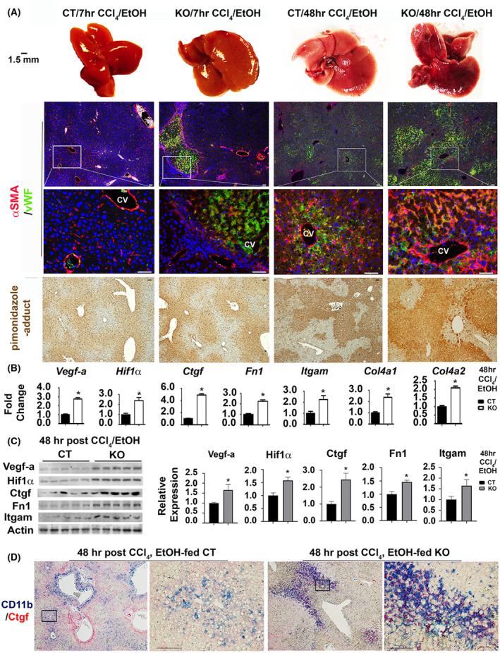
Yap1 deficiency in hepatocytes enhances hypoxia, vascular remodeling, and infiltration of CD11b+ inflammatory cells into Ctgf enriched microenvironments after ethanol/CCl4 treatment. (A) Macroscopic visualization shows hemorrhage in ethanol‐fed Yap1 KO livers at 48 h post CCl4 intoxication (first row). Vascular endothelial cells and activated hepatic stellate cells were stained with vWF and αSMA (second and third rows). Hypoxia was stained based on pimonidazole‐adducts (fourth row). Scale bar: 100 μm. Q‐RT‐PCR analysis (B) and Western blotting (C) detected upregulation of gene signatures for hypoxia and vascular remodeling. Values in (B) represent means ± SD from five animals. *p < .05. Relative levels of Vegf‐a, Hif1α, Ctgf, Fn1, and Itgam proteins in KO in comparison to CT groups (n = 5 per group) were quantified based on densitometrical analyses of band intensities in Western analyses and were expressed as means ± SD. *p < .05. (D) Dual staining showed extensive infiltration of CD11b+ macrophages into Ctgf enriched microenvironments in the damaged Yap1 KO livers. Scale bar: 100 μm
