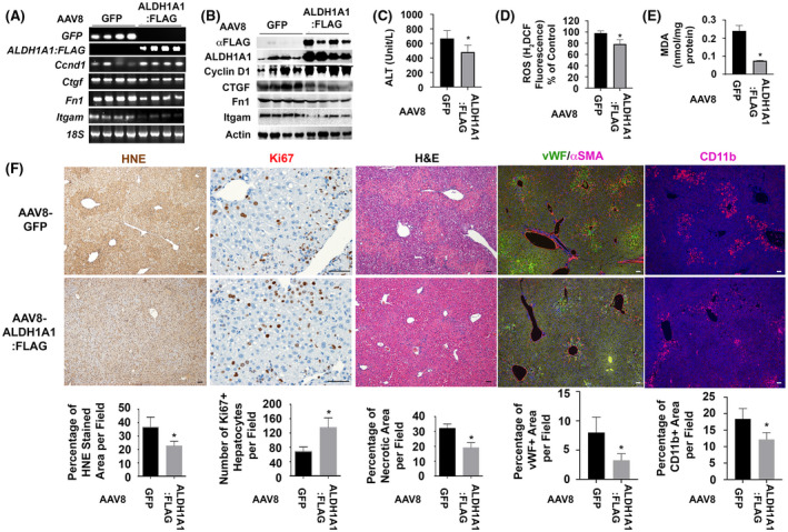
FIGURE 6.

Ectopic expression of ALDH1A1 reduces oxidative stress, decreases hypoxia, attenuates vascular remodeling, ameliorates hepatic inflammation, and enhances hepatocyte proliferation during ethanol/CCl4–induced liver damage. (A and B) RT‐PCR analysis and Western blotting detected AAV8‐ALDH1A1:FLAG or AAV8‐GFP in transduced ethanol‐fed livers at 48 h post CCl4 intoxication. Ectopic expression of ALDH1A1 was associated with upregulation of Cyclin D1 and downregulation of Ctgf, Fn1, and Itgam at mRNA and protein levels. (C–E) Reduction of serum ALT (C), hepatic MDA (D), and ROS (E) were observed in the EtOH/CCl4‐damaged AAV8‐ALDH1A1:FLAG‐transduced livers. Values in (C–E) represent means ± SD (n = 5 mice per group). *p < .05. (F) IHC showed that ectopic expression of ALDH1A1 enhanced number of proliferating hepatocytes (Ki67 staining) and caused reduction in lipid peroxidation (HNE staining), hepatic necrosis (H&E staining), vascular remodeling (αSMA/vWF staining), and inflammation (CD11b staining) during ethanol/CCl4‐induced liver injury. Values were means ± SEM based on quantification of images from more than 10 fields (200× magnification) from 5 mice per group. *p < .05
