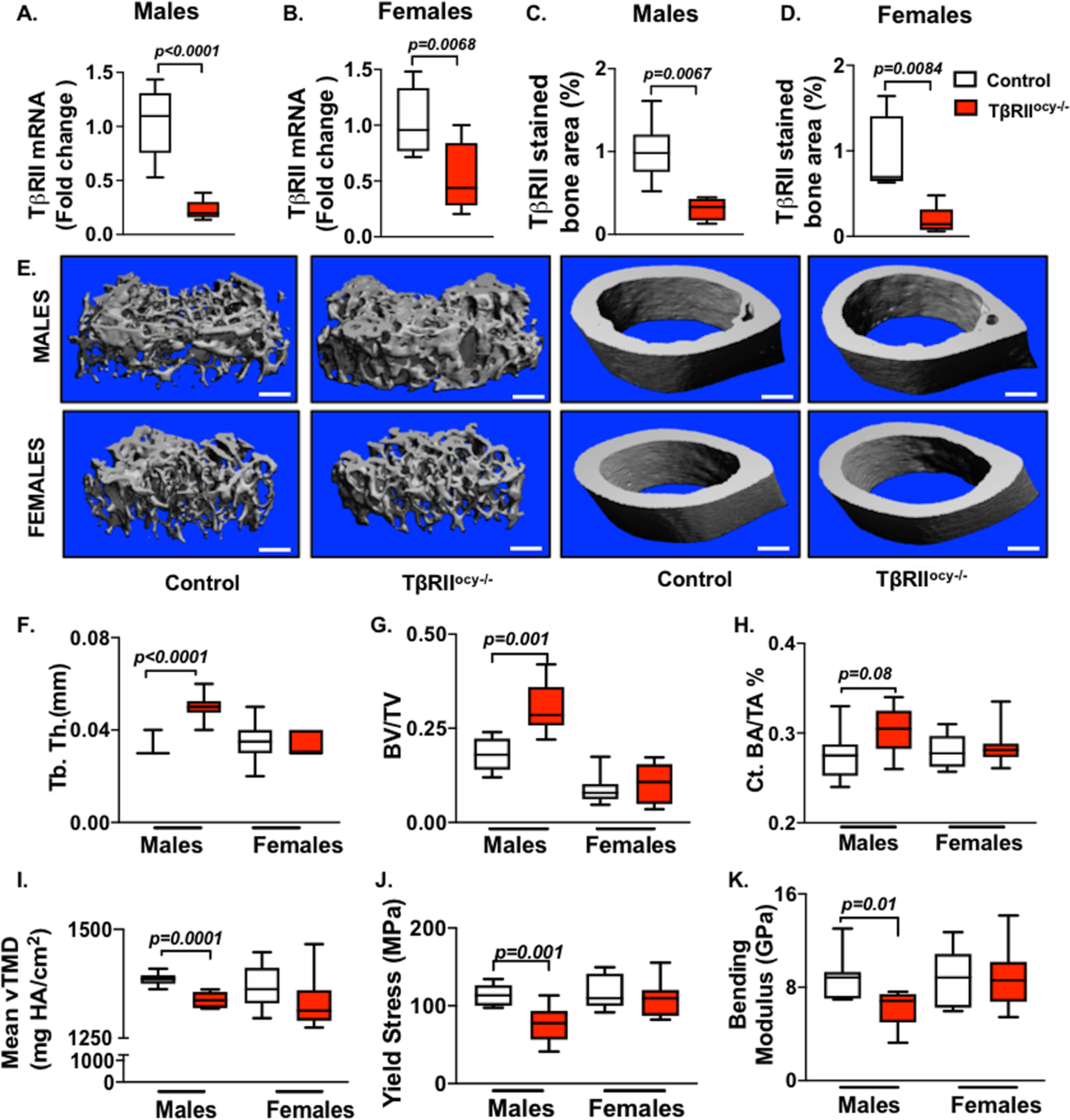
Figure 1. Loss of osteocyte-intrinsic TGFβ signaling impacts bone mass and quality in male mice but does not affect female mice.

In male and female TβRIIocy−/− mice, knockdown of TβRII at mRNA (A, B) (n=6–9 mice/ group) and protein (C, D) (n=4–6 mice/ group) level was assessed. μCT analysis was conducted on femurs from control and TβRIIocy−/− male and female mice (15-week-old). Representative μCT reconstructions of trabecular (left) and cortical (right) bone (E) along with the trabecular bone thickness (Tb. Th.) (F) and volume (BV/TV) (G), and cortical bone area (BA/TA) (H) and mineralization (Ct. Min) (I) are shown (n=7–8 mice/group). Flexural testing of femurs from male and female control and TβRIIocy−/− mice shows bone quality parameters yield stress (J) and bending modulus (K) (n = 7 mice/group). Data for A-D are presented as mean ± SEM and as mean ± SD for E-K and statistically significant difference were determined with Student’s t test. Scale bar for E is 100μm (n = 7–8 mice/group).
