
Figure 2. Osteocyte-specific disruption of TβRII reduces PLR genes in male but not female mice.
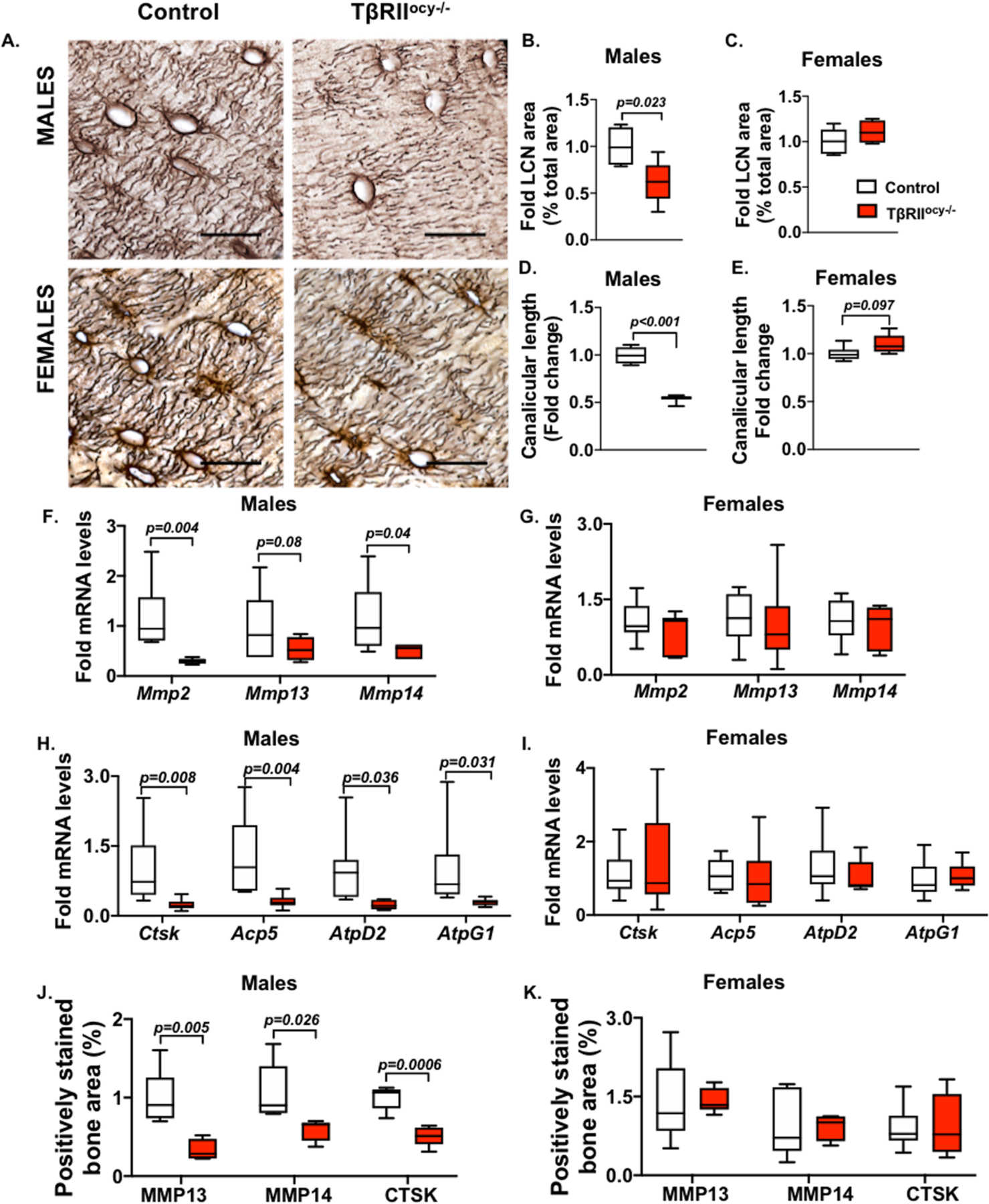
15-week-old male and female control and TβRIIocy−/− mouse bones were processed for mRNA and protein. Osteocyte lacuno-canalicular network (LCN) in the femoral cortical bones was assessed by Ploton stain. Representative images (A) and quantification of LCN area (B, C) and canalicular length (D, E) are shown. Scale bar, 20μm; n=4–6 mice/group and 4 ROI/mouse. Expression of PLR genes Mmp2, Mmp13 and Mmp14 (F, G) and Ctsk, Acp5, Atp6v0d2 (AtpD2) and Atp6v1g1 (AtpG1) are shown (H-I) (n=6–9 mice/ group). Immunohistochemistry (IHC) for MMP13, MMP14, and CTSK on femoral cortical bones of male and female control and TβRIIocy−/− mice shows percent positively stained bone area (J- K) (n =3–4 mice/group and 4 ROI/mouse). Scale bar, 30μm. Error bars indicate mean ± SEM, statistically significant differences were calculated with Student’s t test.