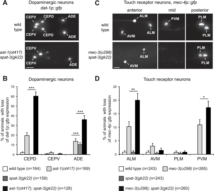
Fig 9. Effect of PRC1 on dopaminergic neurons and touch receptor neurons.
(A) Expression of the dopaminergic marker (dat-1p::gfp, vtIs1) in the head of an early larva. In wild type animals expression is observed in the 6 dopaminergic neurons of the head (2 CEPV, 2 CEPD, 2 ADE). In ast-1(ot417); spat-3(gk22) double mutants expression is lost in some CEPD and ADE (expression lost in the two CEPD and one of the ADE in this example). Dorsal views, anterior is left, scale bar = 5 μm. (B) Percentage of early larvae that display for each class of head dopaminergic neurons a loss of dat-1p::gfp (vtIs1) expression in at least one neuron (error bars show standard error of proportion, n = number of animals analyzed, *** p<0.001, Fisher’s exact test). (C) Expression of the touch receptor neuron marker (mec-4p::gfp, zdIs5) in the anterior, mid and posterior regions of late larvae (L4). In wild type animals expression is observed in the 6 touch receptor neurons (2 ALM, 1 AVM, 2 PLM, 1 PVM). In mec-3(u298); spat-3(gk22) double mutants expression is lost in some ALM and PVM (expression lost in one of the two ALM and in PVM in these examples). Lateral views, anterior is left, scale bar = 5 μm. (D) Percentage of late larvae (L4) that display for each class of touch receptor neurons a loss of mec-4p::gfp (zdIs5) expression in at least one neuron (error bars show standard error of proportion, n = number of animals analyzed, * p<0.05, ** p<0.01 Fisher’s exact test).

