
FIGURE 3.
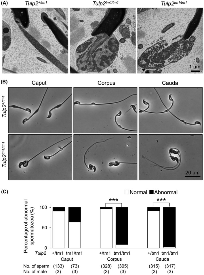
Observation of sperm morphology in the epididymis. (A) Observation of spermatozoa in the cauda epididymis with transmission electron microscopy (TEM). The midpiece was disrupted in Tulp2tm1 / tm1 mice. (B) Representative images of spermatozoa collected from three sections (caput, corpus, and cauda) of epididymis. (C) Percentages of abnormal spermatozoa collected from the three sections of epididymis