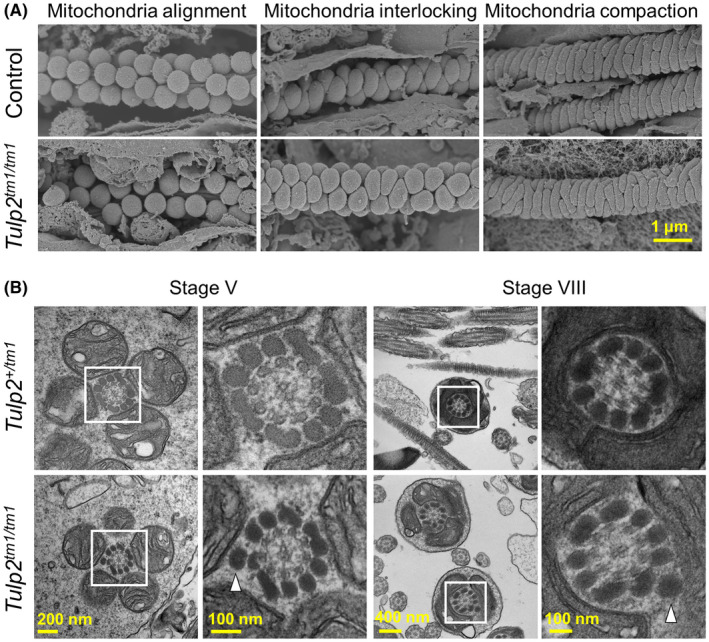
FIGURE 4.

Observation of sperm morphology in the testis. (A) Formation of the mitochondrial sheath during spermatogenesis observed with scanning electron microscopy (SEM). Spherical mitochondria align around the axoneme (left), change their shape in the mitochondrial interlocking step (middle), and form the mitochondrial sheath in the mitochondrial compaction step (right). Wild‐type (WT) and Tulp2 +/ tm1 mice were used as controls. (B) Observation of spermatids with transmission electron microscopy (TEM). Abnormal extra outer dense fibers (ODFs) are highlighted with white arrowheads. Higher magnification images of the boxed areas are shown to the right
