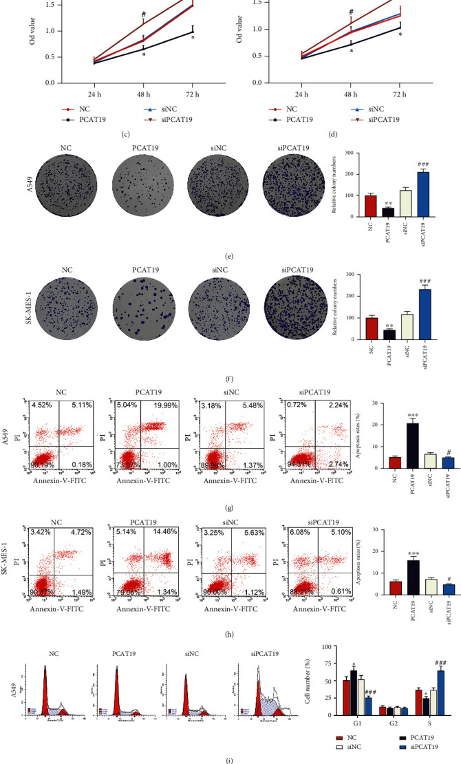
Figure 3.

Upregulated PCAT19 inhibited LC cell proliferation while promoting LC cell apoptosis. (a, b) The expression of PCAT19 in A549 and SK-MES-1 cells transfected with the PCAT19 or siPCAT19 vector was detected by qRT-PCR. (c, d) The viability of A549 and SK-MES-1 cells transfected with the PCAT19 or siPCAT19 vector was detected by the MTT assay. (e, f) Colony formation assay was conducted to observe the growth of A549 and SK-MES-1 cells transfected with the PCAT19 and siPCAT19 vector. (g, h) Apoptosis of A549 and SK-MES-1 cells transfected with the PCAT19 or siPCAT19 vector was detected by flow cytometry. (i, j) Flow cytometry was used to detect the cycle of A549 and SK-MES-1 cells transfected with the PCAT19 or siPCAT19 vector. ∗P < 0.05, ∗∗P < 0.01, and ∗∗∗∗P < 0.001 vs. NC; #P < 0.05, ##P < 0.01, and ###P < 0.001 vs. siNC. Abbreviations: LC: lung cancer; qRT-PCR: quantitative real-time polymerase chain reaction; MTT: methylthiazolyldiphenyl-tetrazolium bromide; NC: negative control.
