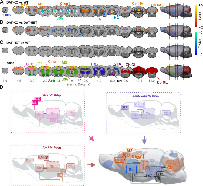
Fig. 2. Brain morphology comparison between DAT-KO, DAT-HET and WT rats and illustration of associative, limbic and motor loops affected by volumetric changes in DAT-KO rats.
A Comparison between DAT-KO and WT rats revealed a significant pattern of decreased relative brain volume in DAT-KO rats (blue scale), with clusters covering predominantly striatum, hippocampus and olfactory bulb. In contrast, relative brain volume in orbitofrontal, cingulate and motor regions as well as in thalamus, midbrain and cerebellum were significantly increased (red scale). B Comparison between DAT-KO and DAT-HET rats revealed a comparable, however less pronounced pattern. C Comparison between DAT-HET and WT rats detected no significant differences between these groups. D Morphological alterations in DAT-KO compared to WT rats demonstrate an overlap with motor, limbic and associative loops (orange—volume gain, blue—volume loss, thresholded at p < 0.01, whole-brain analysis). For visualization purposes, all results in (A–C) are thresholded at a cluster-defining-threshold level of p < 0.01 and only clusters with a size larger than 500 voxels are plotted. White contours signify regions surviving threshold-free cluster enhancement with voxel-wise whole-brain family-wise error correction at p < 0.05. Acb accumbens, Cb cerebellum, Cb I-IV cerebellar lobules I-IV, Cb lat. cerebellum, lateral part, Cb GL granular layer of the cerebellum, Cb ML molecular layer of the cerebellum, Cing1 cingulate cortex area 1, dStr dorsal striatum, HC hippocampus, M1 primary motor cortex, M2 secondary motor cortex, OFC orbitofrontal cortex, OlfB olfactory bulb, SN substantia nigra, STN subthalamic nucleus, Th thalamus, VTA ventral tegmental area.

