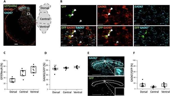
Figure 2.
The AAV2-mDlx enhancer primarily expressed in GAD65-containing neurons in the lumbar spinal cord. (A) Sample tissue section and illustration of the spinal cord showing a distinct layer of GAD67-containing synaptic terminals and neurons (cyan) whereas GAD65-containing neurons are more dispersed (red). The AAV2-mDlx-driven viral expression is shown in green (GFP). (B) A highlighted section (panel A) indicates co-localization of GFP and GAD65, but not GAD67. There are also instances of GAD65+ cells where GFP was not expressed (dim arrow). (C,D) Fraction of co-localization of GFP+ cells and cells containing NeuN (C, N = 2 rats, n = 3 sections/rat) and GAD65 (D, N = 3 animals, n = 3 sections) in the three dorso-ventral regions. (E) The dense layer of GAD67-containing neurons in the dorsal horn (substantia gelatinosa) has little or no overlap with GFP. (F) Cell count (shown as %) of the co-expression in the three regions between GAD67 and GFP positive cells (N = 3 animals, n = 3 sections/animal).

