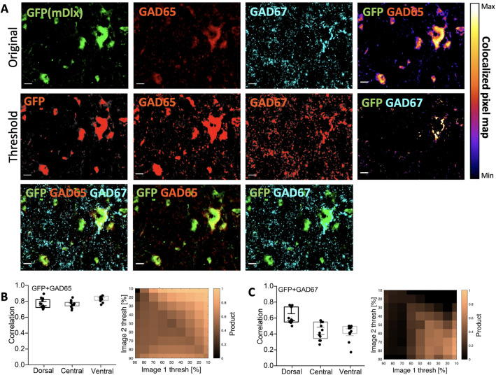
Figure 3.
AAV2-mDlx has expression in GABAergic neurons, specifically the GAD65+ neurons. (A) Top row: original images of mDlx reporter (GFP, green) and GAD65 (red) and GAD67 (cyan) and a colocalization pixel map of GFP/GAD65 (right). Middle row: thresholded versions of top row, with the overlap in heat map (right) (scale bar 20 m). Bottom row: overlap of GFP/GAD65/GAD67, GFP/GAD65 and GFP/GAD67, respectively. (B,C) Varying the threshold and calculating the co-localization as correlation (C). Note N = 4 animals, n = 3 sections/animal.

