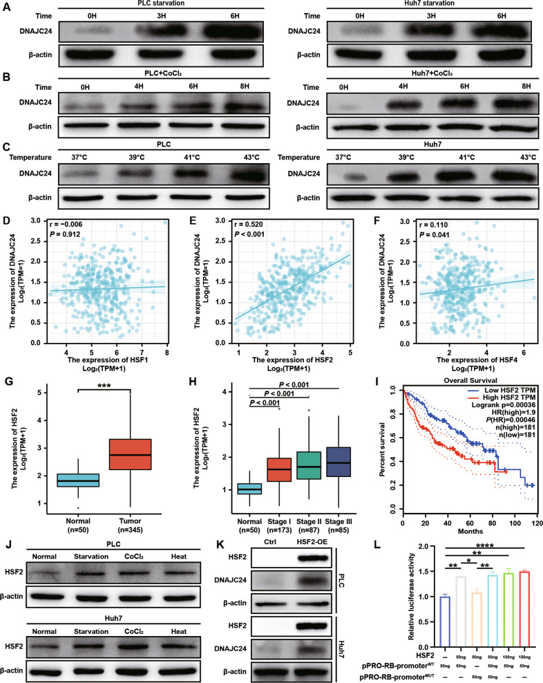
Fig. 3. External stimuli such as starvation, hypoxia and heat upregulate DNAJC24 expression through HSF2 in HCC cells.
A The cell culture medium of PLC and Huh7 was changed to PBS and the cells were cultured in PBS for 0h, 3h and 6h respectively and then the cells were lysed. Western blotting was performed to determine levels of DNAJC24 protein. B Adding CoCl2 (final concentration 200 μM) to the medium of PLC and Huh7 cells to simulate hypoxic environment, and the cells were incubated in it for 0h, 4h, 6h, 8h respectively and then lysed. Western blotting was performed to determine levels of DNAJC24 protein. C PLC and Huh7 cells were cultured at 37 °C, 39 °C, 41 °C, 43 °C respectively in 5% CO2 for 3h, and then the cells were lysed. Western blotting was performed to determine levels of DNAJC24 protein. D–F Analysis of the possible correlation between DNAJC24 and HSF1 (D), HSF2 (E), HSF4 (F) mRNA levels based on the data from the TCGA HCC dataset. Data were analyzed using Spearman’s rank correlation coefficient. G Box plots of HSF2 expression in cancer and paired normal liver tissues of HCC patients based on data from the TCGA HCC dataset. H Box plots of HSF2 expression in normal liver and tumor tissues (Clinical stages I–III) based on data from the TCGA HCC database. I The Kaplan–Meier survival analysis of overall survival of HCC patients analyzed by GEPIA (top 50%, high; bottom 50%, low). J Lysis of PLC and Huh7 cells after applying starvation, hypoxia, and heat (40 °C) stimulation to 3h. Western blotting was performed to determine levels of HSF2 protein. K Stable cell lines with HSF2 overexpression were constructed using lentiviral transfection in PLC and Huh7 cells, and the protein levels of HSF2 and DNAJC24 were detected by Western blotting after cell lysed. L DNAJC24 wild-type promoter (pPRO-RB-promoterWT) and mutant promoter (pPRO-RB-promoterMUT)were ligated to the reporter vector. A dual luciferase assay was performed to assess the effects of HSF2 overexpression on DNAJC24 transcription in PLC cells. Data were presented as mean ± SEM. n = 3. *P < 0.05, **P < 0.01, ***P < 0.001, ****P < 0.0001.

