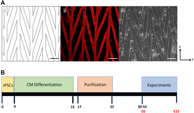
FIGURE 1.
(A) Chevron pattern of 30 µm lanes connected at 15°: (i) CAD drawing of the pattern; (ii) Matrigel stained with laminin; (iii) Pattern seeded with control CPVT stem-cell derived cardiomyocytes. Scale bars are 100 µm. (B) Timeline of cell culture, differentiation, purification, and experiments. Day 0 denotes the start of CM differentiation and E0 denotes the start of experiments after cells are seeded onto a micropatterned substrate.

