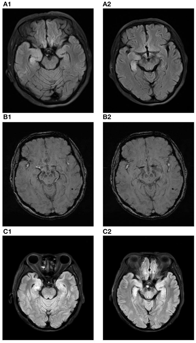
Figure 1.

(A1,A2) show the increased volume of the right hippocampus and hyperintensities in the fluid attenuated inversion recovery (FLAIR) sequence at the time of initial diagnosis. (B1,B2) show microbleed in the left temporal lobe in the susceptibility weighted imaging (SWI) sequence at the time of initial diagnosis. (C1,C2) show the increased volume of the right hippocampus and hyperintensities in the fluid attenuated inversion recovery (FLAIR) sequence ten months after diagnose.
