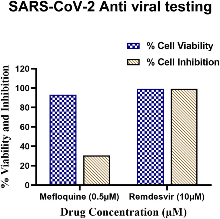
Figure 10.
Antiviral testing of Mefloquine against SARS-CoV-2 in VeroE6 cells. (i) The ~104 VeroE6 cells were pre-treated with Mefloquine and Remdesivir drug for 30 h at 37°C. Post-incubation, cells were stained with Hoechst 33342 and Sytox orange fluorescent dyes. The drug and control molecule treated cells were counted through stained Hoechst and Sytox image by using MetaXpress software. From raw images, the % average cell viability with control cells were calculated and the bar diagram highlighted in blue colour was plotted. (ii) 1 MOI of SARS-CoV-2 virus infected Vero E6 cells were treated with mefloquine drugs. The cells were fixed, permeabilized, and stained with mAb specific to SARS-CoV-2 nucleocapsid (primary) followed by alexafluor 568-labeled antibody (secondary) and all the cells were stained with Hoechst 33342 stain. The nucleocapsid positive and total nuclei cells were counted from the stained image and the % average cell inhibition with all the controls were calculated and the bar diagram highlighted in brown colour. All these assays were performed in triplicate wells.

