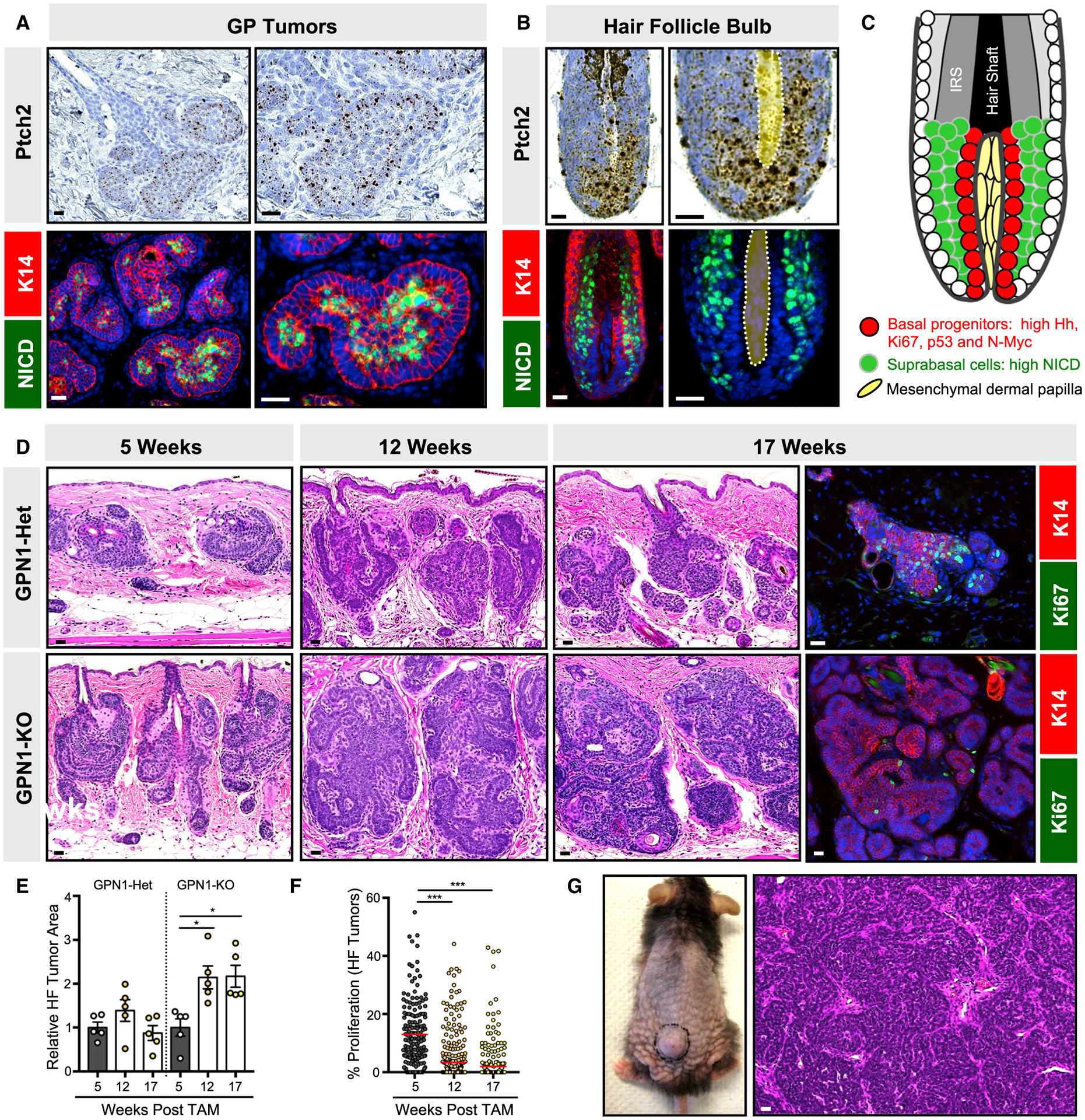
Figure 3. Notch1-deficient microscopic tumors do not undergo spontaneous regression.

(A) GP tumors have elevated Hh target genes, as assessed by Ptch2 mRNA, in peripheral basal cells (top panels) and high NICD (green) in interior suprabasal cells (lower panels). Red, K14.
(B) In the growing anagen hair follicle, basal matrix progenitor cells directly abutting the dermal papilla (yellow, dotted) express high Ptch2 (top panels), whereas their suprabasal progeny express high NICD (green, bottom panels).
(C) Schematic for basal progenitors (red) and suprabasal progeny (green) in the hair follicle bulb. IRS, inner root sheath.
(D) Histology of HF-derived GP tumors that are either Notch1-heterozygous (GPN1-Het) or deleted (GPN1-knockout [KO]), 5–17 weeks post-TAM. Right panels, tumor proliferation as assessed by Ki67 (green), 17 weeks post-TAM.
(E) Quantitation of GPN1-Het and GPN1-KO tumor area.
(F) Quantitation of GPN1-KO tumor proliferation.
(G) Photo and histology of GPN1-KO macroscopic tumor.
Data are represented as mean ± SEM, with significance calculated by one-way ANOVA. Significance for beeswarm plots was calculated using a linear mixed model. *p < 0.05; ***p < 0.001. Scale bars, 50 μm. See also Figure S3 and Table S1 for animal numbers.
