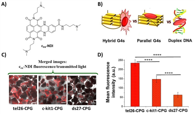
Figure 8.
A) Chemical structure of cex‐NDI. B) Schematic picture of the fluorescent behaviour of cex‐NDI when bound to hybrid G4s, producing a dramatic fluorescence enhancement, and parallel G4s or duplex DNA with a weaker fluorescence emission. C) Representative confocal images of tel26‐, c‐kit1‐ and ds27‐functionalized CPG supports after incubation with the cex‐NDI. Merged images of cex‐NDI fluorescence and transmitted light. Scale bars correspond to 100 μm. D) Mean fluorescence intensity values (±S.D.) taken from different edge glass beads‐containing regions of each image acquired for tel26‐, c‐kit1‐ and ds27‐functionalized CPG supports. p‐values have been calculated using the Student's t‐test (****p<0.0001). Adapted with permission from Ref. [41] Copyright 2018, Elsevier.

