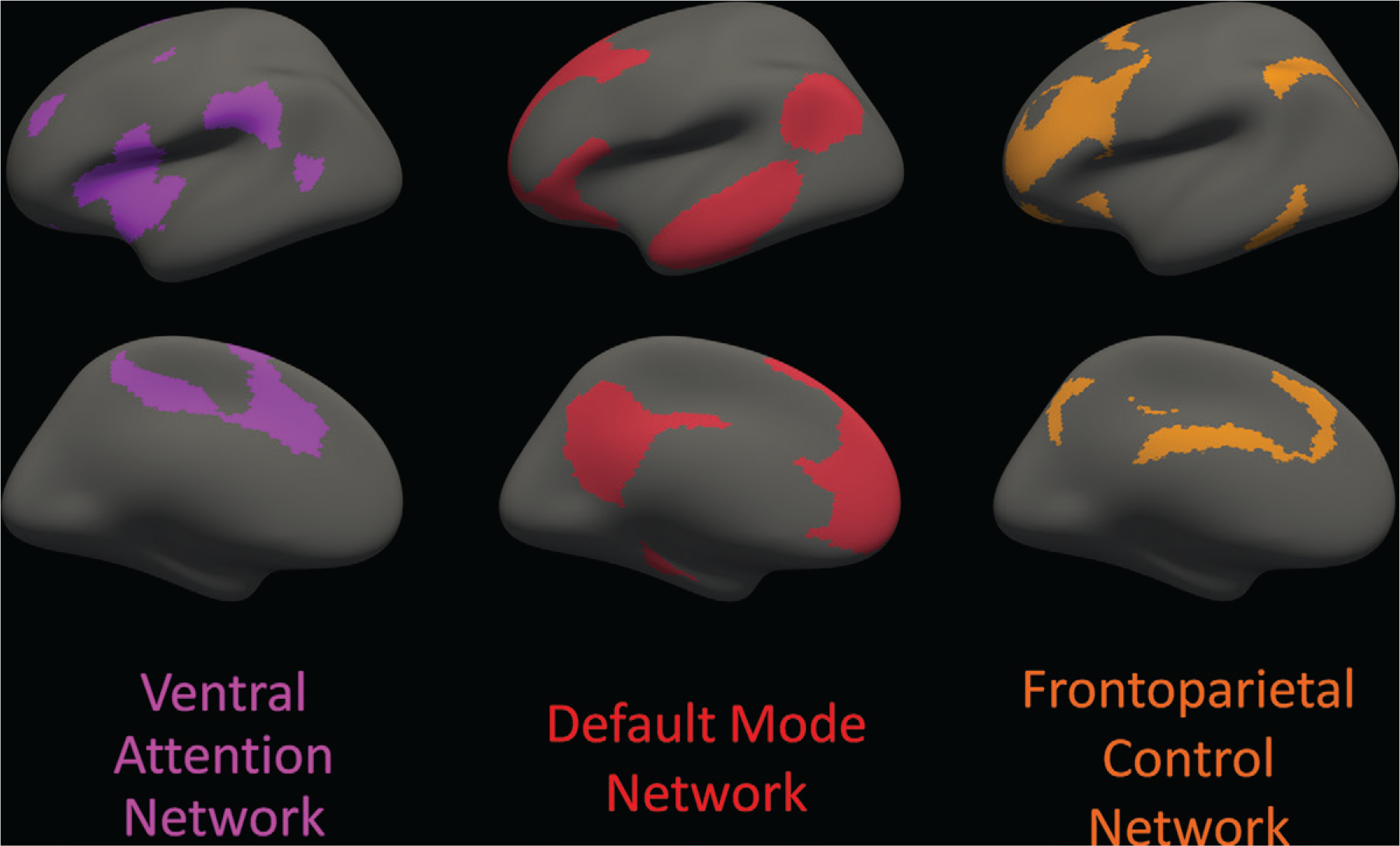
Figure 1:

Cortical areas involved with each of the three distributed cortical association networks analyzed are displayed on an inflated brain surface, viewed from the lateral aspect (top row) and the medial aspect (bottom row) of the left hemisphere; the right hemisphere (not shown) is nearly a mirror image of the left hemisphere. The regions are based on a standard functional parcellation of the human brain from a resting-state functional MRI study of 1000 healthy individuals. The three specific networks of interest are the ventral attention network (purple), default mode network (red), and frontoparietal control network (orange). Cortical regions in the ventral attention network include portions of the middle frontal gyrus, inferior frontal gyrus (pars triangularis and pars opercularis), insula, supramarginal gyrus, posterior superior temporal sulcus, superior frontal gyrus, anterior and posterior cingulate, and precuneus. Cortical regions in the default mode network include portions of the medial prefrontal cortex; precuneus; posterior cingulate; parahippocampal gyrus; superior, middle, and inferior temporal gyri; and inferior parietal gyrus. Cortical regions in the frontoparietal control network include portions of the superior and middle frontal gyri, inferior temporal gyrus, supramarginal gyrus, anterior and posterior cingulate, and precuneus. Note that some of the gyral-based parcellated areas are components of more than one network defined by the functional parcellation. Source.–Reference 15.
