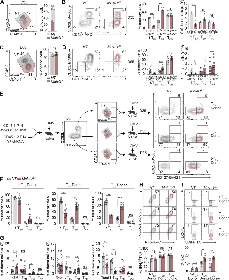
Figure 2.
Malat1 regulates CD8+ T cell memory formation and represses generation of secondary TEM cells. (A and C) Quantification of splenic NT and Malat1KD ratios on days 35 and 65 after infection. (B and D) Representative flow cytometry plots of t-TEM, TEM, and TCM cells (left) and quantification (right) among co-transferred cells. (E) P14 CD8+ T cells were transduced with Malat1 shRNA (Malat1KD, CD45.1) or nontarget shRNA (NT, CD45.1.2) and adoptively co-transferred at a 1:1 ratio into CD45.2 recipient mice, which were then infected with LCMV. 35 d after primary infection, Malat1KD and NT cells were sorted from t-TEM, TEM, or TCM subsets, mixed at a 1:1 (5,000 Malat1KD cells/5,000 NT cells) ratio, and adoptively transferred into naive CD45.2 recipient followed by infectious challenge with LCMV (secondary infection). (F) Frequency of secondary memory populations derived from transferred t-TEM (left), TEM (middle), and TCM (right) donor cells was assessed at 30 d after secondary LMCV infection. (G) Quantification of secondary memory subsets derived from t-TEM (left), TEM (middle), and TCM (right) donor populations. (H and I) Malat1KD and NT secondary t-TEM, TEM, and TCM cells were cultured ex vivo in the presence of cognate gp33-41 peptide for 5 h and frequency of IFNγhiTNFhi (H) or IL-2+ (I) cells measured. All data are from one representative experiment out of two independent experiments with n = 4–7 (A–D), n = 9–10 (E–G), or n = 4–6 (H and I) mice per group; *, P < 0.05; **, P < 0.005; ***, P < 0.0005, paired t test. Graphs indicate mean ± SEM, symbols represent individual mice. D, day.

