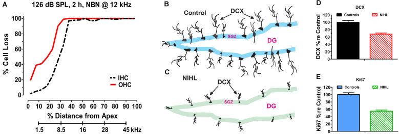
Figure 2.
Noise-inducedhearing loss suppresses hippocampal cell proliferation andneurogenesis. (A) Cochleogram showing massive loss of outerhair cells (OHC) and inner hair cells (IHC) in the noise-exposed cochlear several months after a 2-h unilateral exposure to narrowband noise (NBN) centered at 12 kHz and presented at 126 dB SPL. Percent cell loss plotted as function of percent distance from the apex of the cochlear. Cochlear place related to frequency using rat tonotopic map on lower abscissa. (B) Schematic of dentate gyrus (DG) of hippocampus from normal control showing immunolabeled doublecortin (DCX) soma in the subgranular zone (SGZ); note extensive immunolabeled processes emanating from soma. (C) Schematic of DG of hippocampus several months after a noised induced hearing loss (NIHL) showing immunolabeled DCX) soma in the subgranular zone (SGZ). Note reduced number of DCX soma and paucity of labeled processes in the NIHL hippocampus compared to normal control (panel B). (D) Schematic showing relative number (% re Control: percentage relative to control) of DCX labeled neurons in hippocampus of normal control rats (100%) and rats with noise-induced hearing loss (NIHL). (E) Schematic showing relative number (% re Control: percentage relative to control) of Ki67 labeled neurons in hippocampus of normal control rats (100%) and rats with noise-induced hearing loss (NIHL).

