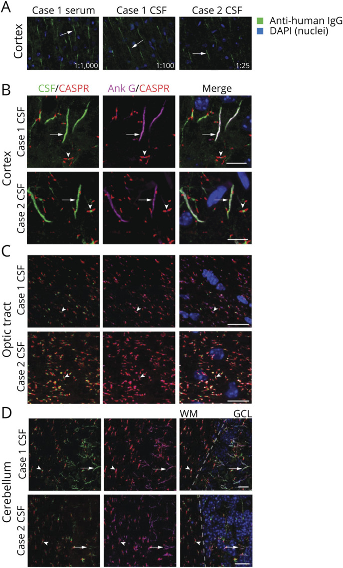
Figure 1. Patient Antibodies Localize to the AIS and NoR.

(A) Mouse brain sections were immunostained with case 1 serum (1:1,000 dilution), case 1 CSF (1:100 dilution), and case 2 CSF (1:25 dilution). In all cases, patient IgG (green) immunostained structures consistent with the AIS (arrows). Images were captured at 20× on an epifluorescent microscope. (B–D) Anatomic coimmunostaining of case 1 and 2 CSF IgG (green) with the AIS and nodal marker AnkG (magenta) in the (B) cortex, (C) optic tract, and (D) cerebellum. In D, the dotted line indicates the boundary between the cerebellar WM and GCL. Arrows indicate axon initial segments while arrowheads indicate nodes of Ranvier which are demarcated by CASPR in red. All scale bars = 10 µm. Images were captured by confocal microscopy at 60 or 100×. AIS = axon initial segment; AnkG = ankyrin G; GCL = granule cell layer; IgG = immunoglobulin G; NoR = node of Ranvier; WM = white matter.
