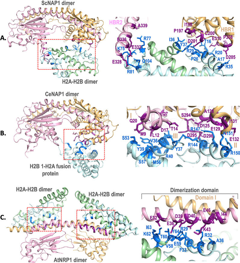
Fig. 2.
Structural depiction of the histone H2A–H2B dimer interacting residues of ScNAP1, CeNAP1 and AtNRP1. NAP dimer is shown as ribbon and monomers are colored pink and tan. Histones H2A and histone H2B are shown as cyan and green ribbon. The interacting residues of NAPs and histones are shown as violet and blue sticks, respectively. The binding site of histones on each NAP is marked with red boxes. A ScNAP1 dimer interacting with H2A–H2B dimer (PDB ID: 5G2E). HBR1, HBR2 and the corresponding interacting residues of ScNAP1 are highlighted. B CeNAP1 dimer interacting with H2B 1-H2A fusion protein (PDB ID: 6K00). Regions I, II, III and the corresponding interacting residues of CeNAP1 are highlighted. C AtNRP1 dimer interacting with H2A–H2B dimer (PDB ID: 7C7X). The dimerization domain I and the interacting residues of AtNRP1 are highlighted. Interacting residues are collated from PDBSUM (www.ebi.ac.uk/thornton-srv/databases/cgi-bin/pdbsum/) and all structural depictions are made using Pymol (www.pymol.org)

