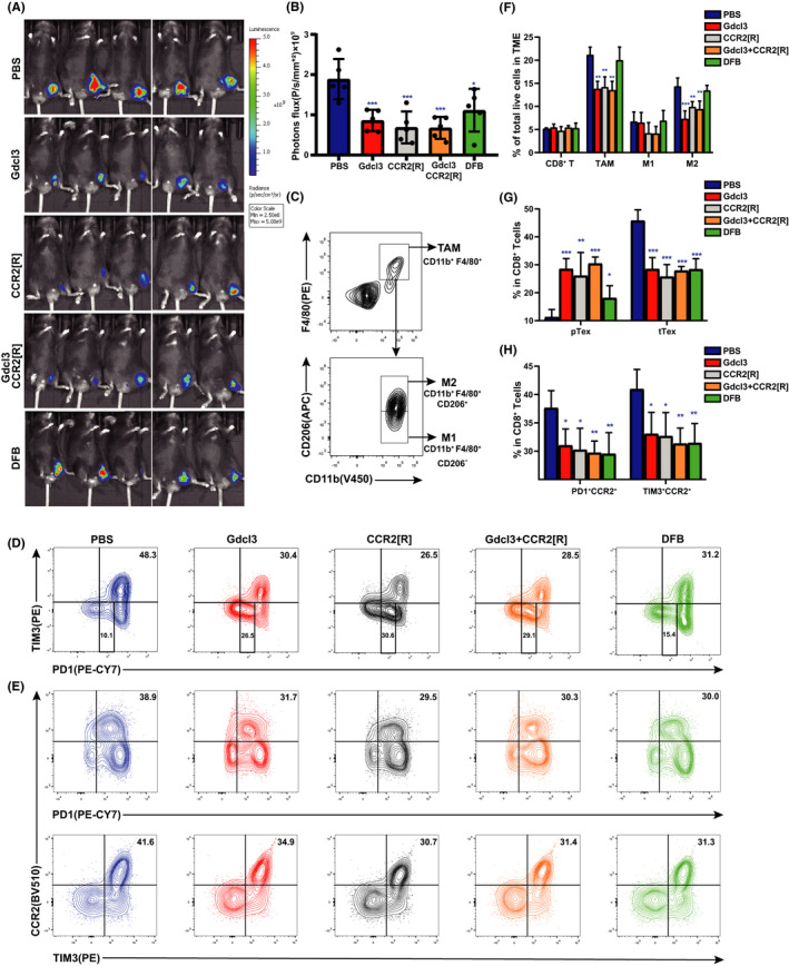
FIGURE 6.

Dahuang Fuzi Baijiang decoction (DFB) inhibits shift from progenitor exhausted T cells (pTex) to terminal exhuasted T cells (tTex) independent of macrophages. (A) In vivo imaging of small animals in the colorectal subcutaneous model. (B) Fluorescence statistics of figure (A). (C) Flow cytometry gating strategies used to define tumor‐associated macrophages (TAM) (CD11b+ F4/80+), M1‐TAM (CD11b+F4/80+CD206−) and M2‐TAM (CD11b+F4/80+CD206+). (D) Representative flow plots and frequency of PD‐1 and TIM3 in tumor infiltration of mice in each group. (E) Representative flow plots and frequency of recombinant chemokine C‐C‐motif receptor 2 (CCR2) on the tumor‐infiltrating CD8+ Tex cells of each group. (F) Relative percentages of the tumor‐infiltrating inflammatory cells, as determined by FACS, in the tumor tissues of mice subjected to various treatments. These include CD8+ tumor‐infiltrating lymphocytes, TAM, M1‐TAM, and M2‐TAM (n = 5 per group). (G) Statistical chart of the expression of tTex and pTex on the tumor‐infiltrating CD8+ T cells of each group of mice (n = 5 per group). (H) Statistical chart of the expression of CCR2 on the tumor‐infiltrating CD8+ Tex cells of each group of mice (n = 5 per group). *P < .05, **P < .01, ***P < .001, compared with C57BL/6J (PBS) group. Data in this figure are depicted as mean ± SEM, with all individual points shown. Ordinary one‐way ANOVA P values are shown. GdCl3, gadolinium chloride
