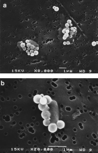
FIG. 2.
Electron micrographs of immunomagnetic beads attached to E. coli O157:H7 cells. Original magnifications: a, ×8,000; b, ×20,000. Cells appear as faint rods with five or more white beads attached. The pores in the polycarbonate nuclear etched membrane can be seen in the background. WD, working distance.

