Figure 3.

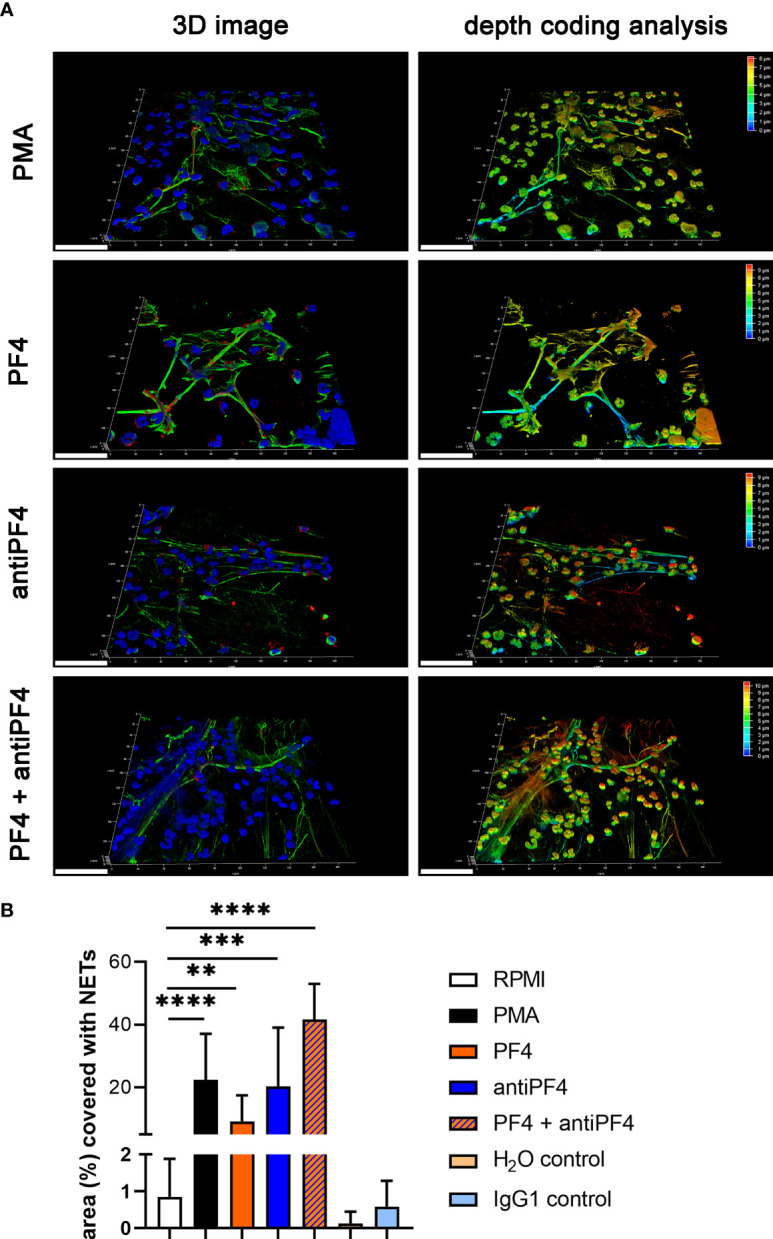
PF4 and antiPF4 are potential NET inducer forming compact NETs. Human blood-derived neutrophils release NETs after 2 h incubation in response to PF4 and antiPF4. NETs were detected by confocal immunofluorescence microscopy. The settings were adjusted to a respective isotype control. (A) Representative 3D reconstructions of z-stacks are presented, showing extracellular DNA-fibers positive for DNA/histone-1 complex (green) and myeloperoxidase (red), (Scale bar = 50 µm). Images were constructed with LAS X 3D Version 3.1.0 software (Leica): PMA = 8.09 µm consisting of 49 sections; PF4 = 9.57 µm consisting of 58 sections; antiPF4 = 9.40 µm consisting of 57 sections; PF4 and antiPF4 = 10.41 µm consisting of 63 sections. Out of this 3D volume a depth coding analysis was conducted and is presented with a depth coding scale bar. (B) In each experiment and for each sample, six randomly taken pictures from two individual slides were analyzed for NET quantification. The area covered with NETs was measured and used for statistic. In one experiment the internal controls H2O and mouse IgG1 were included as control for PF4 (dissolved in H2O) and antiPF4 (mouse IgG1 anti human PF4). Data were analyzed with one-tailed unpaired Student’s t-Test calculated always to negative control (RPMI) (**p < 0.01, ***p < 0.001 and ****p < 0.0001). Data are presented with mean ± SD.
