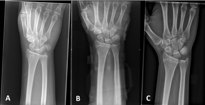
Figure 4.
Radiographs demonstrating a radial styloid fracture of the right wrist at presentation to the emergency department. (A). Radiograph at the 6-week follow-up with the 3D-printed cast. Note that the fracture is not concealed, thus allowing for ideal follow-up. (B) Radiograph at 3 months showing union of the fracture with no loss of reduction. (C)

