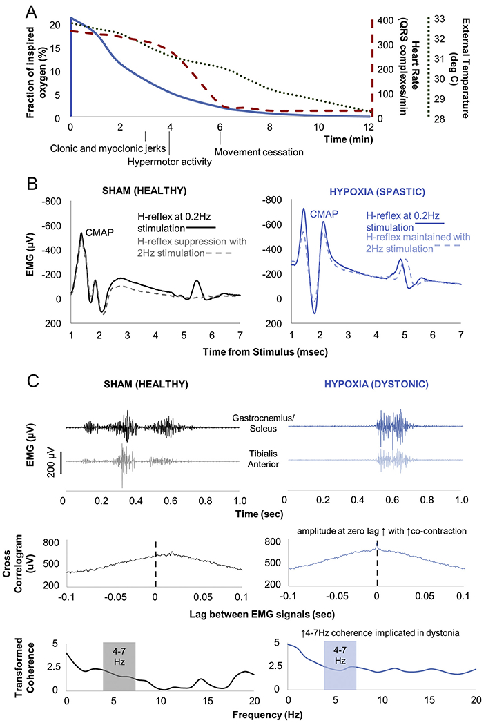
Fig. 1.

Overview of hypoxia exposure and motor electrophysiologic assessments. A) Vital sign and motor changes during neonatal hypoxia exposure. Axes and the vital signs to which they correspond have comparably coded lines. Vital signs did not vary significantly between pups during hypoxia exposure. B) Example Hoffman (H) reflexes. H-reflexes are analogues of the spinal stretch reflex that emerge following compound muscle action potentials (CMAP). H-reflexes normally suppress with high frequency stimulation (sham animal), but do not suppress with upper motor neuron injury (hypoxia-exposed animal). C) Example dystonia measures. Electromyography (EMG) during restrained isometric voluntary hindlimb contraction shows antagonist muscle co-contraction in the sham-exposed animal, but greater co-contraction in the hypoxia-exposed animal. Cross-correlogram measures EMG signal correlation across time. Transformed coherence measures oscillatory synchrony between signals. EMG signals have been rectified and leveled to remove amplitude information. Cross-correlogram and coherence distributions have been normalized using the Fisher z-transform.
