Figure 4.
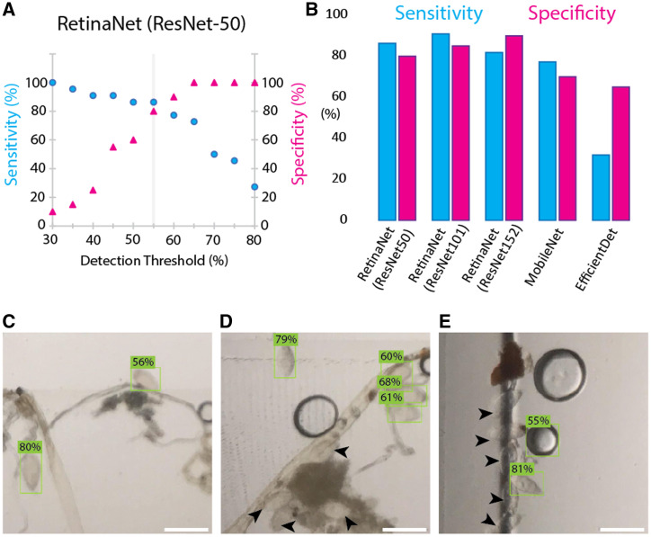
Automated Schistosoma haematobium egg detection using transfer learning. (A) By varying the detection threshold for a single region of interest, the sensitivity (blue dots) and specificity (magenta triangles) of the algorithm as a whole can be tuned. The example is shown for RetinaNet (ResNet-50) with the optimal detection threshold at 55%. (B) The ability of different neural networks to detect the presence of schistosome eggs in an image is quantified using sensitivity and specificity, with expert counts as the ground truth. (C and D) The algorithm performs well at detecting isolated eggs and rejecting other debris from a crowded field of view but does not identify eggs that are clustered with debris (black arrowheads). (E) At the edges of the cartridge, many eggs are not identified by the algorithm. In this image, an air bubble is incorrectly classified as an egg, albeit with lower probability. All scale bars = 200 µm.

