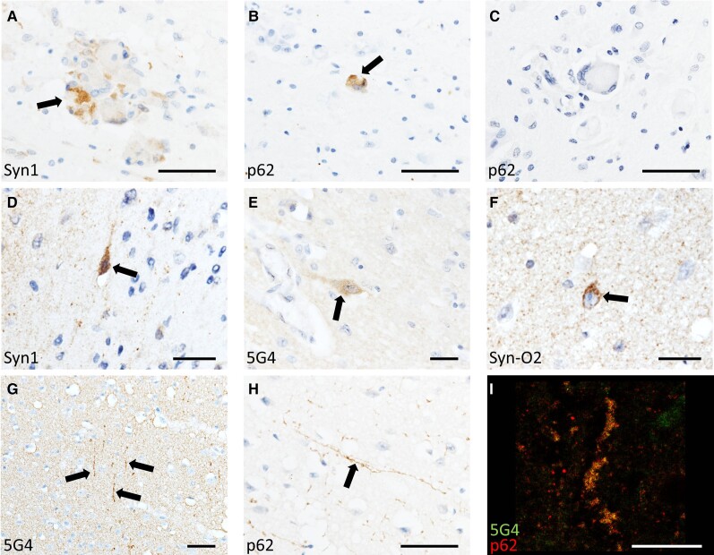
Figure 1.
α-Synuclein in Krabbe disease. Photomicrographs of Krabbe disease (KD) cases demonstrating α-synuclein-immunoreactive globoid cells (A) that were variably positive for the autophagic marker p62 (B and C). Occasional neurons manifested punctate α-synuclein staining (D–F) and neurites that were immunoreactive for α-synuclein and p62 were variably present throughout the cortex of all cases (G
I). Scale bars = 50 µm (A–C, G and H) 25 µm (D–F) and 20 µm (I).

