Figure 2.
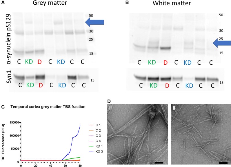
Western blot and RT-QuIC of Krabbe disease brain tissue lysates. The TEAB-soluble fraction of KD temporal cortex grey matter lysates contained pS129 bands at ∼40 kDa (blue arrow) that were not observed in control cases or the DLB case (A). In white matter samples, KD cases had additional bands of pS129 at ∼20 kDa (blue arrow) that were not present in control or DLB cases (B). RT-QuIC of the TBS-soluble tissue fractions demonstrated that KD1 and KD3 grey matter gave positive reactions, defined as Thioflavin-T fluorescence >5 times the control standard deviation, at ∼55 h while no reaction was observed in white matter samples (C). Transmission electron microscopy of RT-QuIC end-products from grey matter of KD1 [D(i)] and KD3 [D(ii)] confirmed the presence of fibrillar structures. It is notable that KD1 RT-QuIC end-products led to elongated fibrils of ∼200–400 nm while KD3 end-products were typically 100–200 nm [E(i) and F(ii)]. Scale bars = 50 nm [D(i and ii)].

