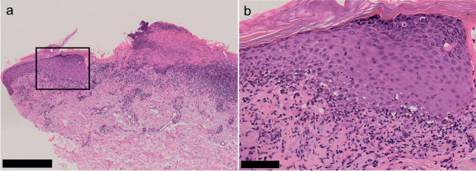
Fig. 2.
Histopathological findings. (a) Hematoxylin-eosin-stained specimens of the erosive papules on the right forearm. Band-like lymphocytic infiltration just beneath the hypertrophic epidermis with compact ortho-hyperkeratosis and hypergranulosis. Focal erosion is shown on the right (scale bar = 500 μm). (b) High magnification of the black square in (a). Vacuolar changes at the dermo-epidermal junction and apoptotic keratinocytes (scale bar = 100 μm).

